Cost-Effectiveness Analysis of 3D Total-Body Photography for People at High Risk of Melanoma

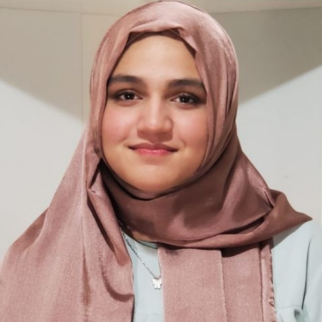
Featured Student: Fatima Iqbal
Fatima Iqbal is a first-year student in the B.A./M.D. program at the University of Missouri–Kansas City. She loves hanging out with friends, baking, and participating in debate tournaments. Her research experiences have strengthened her understanding of how innovation can address challenges in healthcare. Fatima is particularly interested in dermatology, with a specific focus on Mohs surgery and its clinical applications. She is eager to pursue research in Mohs surgery to better understand its role in skin cancer treatment. Fatima hopes to integrate her passion for advocacy and medicine to improve equitable healthcare outcomes on both clinical and policy levels.
Introduction
Melanoma is a fast-growing form of skin cancer that’s especially dangerous when detected late. For patients who are considered high-risk like those with a personal history of melanoma or many atypical moles consistent and accurate skin monitoring is essential. But routine skin exams and even 2D photography can miss early changes.
A recent study published in JAMA Dermatology (March 2025) by Lindsay explored whether 3D total-body photography (3D TBP) combined with sequential digital dermoscopy imaging (SDDI) could improve melanoma surveillance outcomes—and whether it’s a cost-eective strategy in the long run. The study blends clinical care, economics, and tech to answer a question that’s becoming more relevant as imaging tools become more advanced.
Methodology
The researchers used a cost-effectiveness model to simulate the long-term impact of three different surveillance methods for high-risk individuals:
- Standard care – regular full-body skin exams.
- 2D TBP with SDDI – traditional flat photography with dermoscopic follow-up.
- 3D TBP with SDDI – an immersive 3D scanning system that maps the skin’s surface, tracks lesions over time, and flags any subtle changes for further evaluation.
The model accounted for patient outcomes over a lifespan and included factors like:
- Number of melanomas detected early
- Reduction in unnecessary biopsies
- Long-term treatment costs
- Patient quality of life, measured in quality-adjusted life years (QALYs)
- Healthcare spending thresholds (specifically, whether the cost per QALY was under $100,000, which is commonly used as a benchmark for value)
Findings
The results showed that 3D TBP with SDDI had the highest upfront costs, mainly due to the technology itself and the setup required. But over time, it actually saved money by catching melanomas earlier, when they’re easier and less expensive to treat. It also reduced unnecessary procedures, like biopsies, that can cause stress, discomfort, and costs for patients.
Here’s the part that stood out: the incremental cost-effectiveness ratio (ICER) for 3D TBP was around $50,000 per QALY, well below the $100,000 threshold, making it a cost-effective strategy for high-risk patients. It also offered more QALYs than the other methods, showing both clinical and economic benefit.
Strengths and Weaknesses
Strengths:
This study was strong in its design. It looked at both outcomes and cost, two things that matter a lot in real-world decision-making. The use of lifetime modeling, along with established cost thresholds, gave a clear picture of how 3D TBP could be applied in actual practice.
Weaknesses:
One of the study’s limitations is that it’s based on modeling, not a randomized clinical trial. That means it relied on previously published data and assumptions about patient behavior, follow-up, and disease progression. Also, the model focused on high-risk patients, so we don’t know yet how these fi ndings would translate to people at average risk or in underserved areas where access to 3D imaging might be limited.
Applications to Future Practice
This study suggests that 3D total-body photography, when paired with SDDI, could be a valuable tool in dermatology especially for patients at high risk of melanoma. As the technology becomes more available, it could change the way providers approach skin cancer checkings. Early detection means better outcomes, fewer invasive procedures, and ultimately, lower costs for patients and the healthcare system.
For students like me, this study was a reminder that medicine doesn’t happen in a vacuum. The best tools are the ones that work and make sense for patients, clinics, and the system as a whole. 3D TBP could be one of those tools, and I’m excited to see how it fi ts into future practice.
Reference
Lindsay, H., et al. (2025). Cost-Effectiveness Analysis of 3D Total-Body Photography for Melanoma Surveillance. JAMA Dermatology. Published March 26, 2025.
https://jamanetwork.com/journals/jamadermatology/fullarticle/2831499


